
Dental Technology In Columbia, SC
Dental technology has introduced innovative advancements over the last few years, making dental appointments quicker and much more thorough. Some of the laborious tasks of dentistry have been simplified and the process for several of these duties has proven more efficient. Technology has already altered our everyday lives at home and in the workplace, making it only a matter of time until modern developments changed how patients perceived a routine dental appointment. Here are the pieces of technology we have in our office.

-
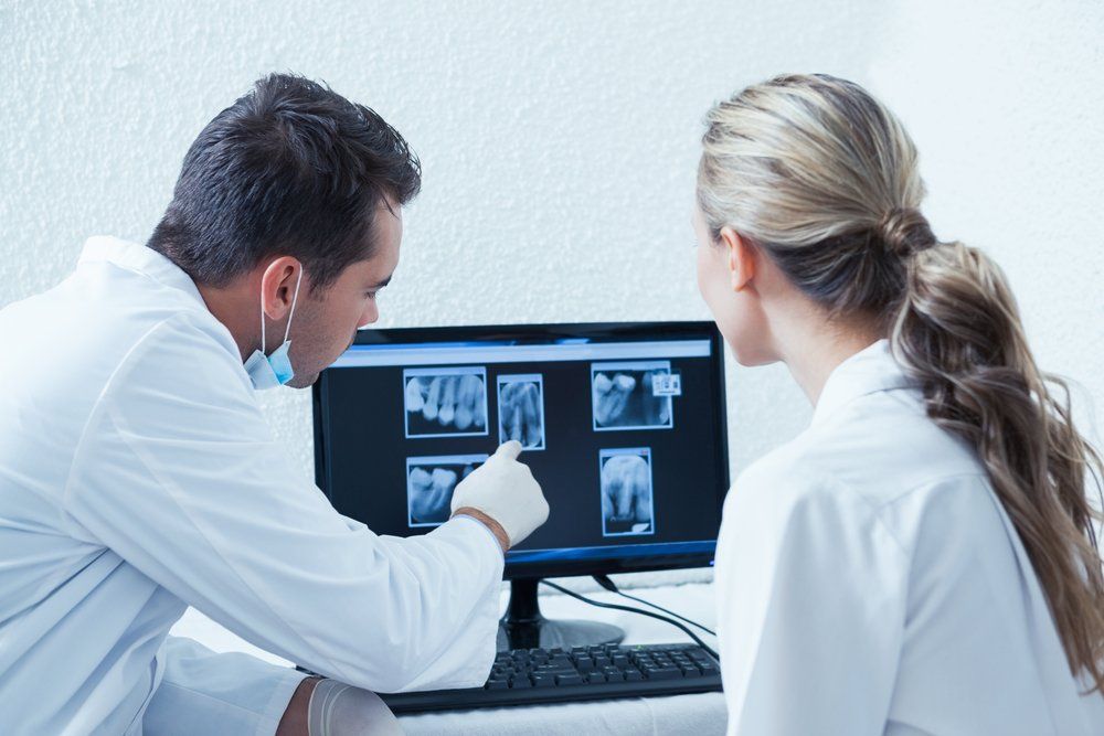
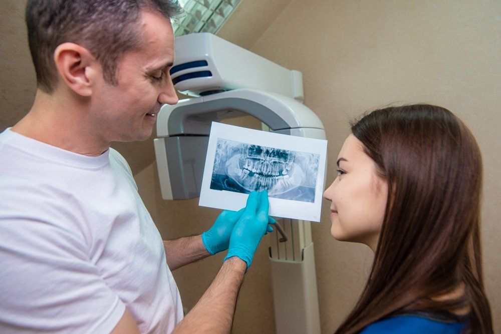
Digital X-Rays
Introduced in 1987, nearly 90 years after traditional x-rays came to fruition, digital radiography combined the power of computer technology with electric sensors and tiny bursts of radiation. Rather than printing the results on film, images form almost as soon as the sensors are placed in our mouths, projecting on a computer screen. Digital x-ray technology does demand additional training for dentists, though the majority of practitioners are adamant that the advantages are worth the commitment. Today, a lot of dental offices only offer patients digital x-rays because, in multiple ways, it is the superior option to traditional radiography.
- Less Expensive | Digital x-rays will generally cost you less than the traditional alternative because the cost of film to develop images for the latter adds up. In contrast, digital x-ray imaging projects right onto our computer.
- Better Storage | Since these digital x-ray images are transferred to a computer system, it allows for easier storage of your oral health records. Your data can be transferred from one dentist to another without any medical data being lost in the exchange.
- Finer Images | Digital x-ray images produce a better resolution than their traditional counterpart. Also, old-fashioned x-rays can only project images in 25 various shades, whereas a digital image can reveal up to 256 shades of grey. Digital radiography also has the advantage of accessing more angles within our mouths, providing a streamlined view of a patient's entire oral structure. With the assistance of computer programs, dentists can even enhance the digital images further, for a focused view.
-
VELscope
VELscope's Vx Enhanced Oral Assessment System is a handheld scope utilized by dentists to help visualize oral tissue abnormalities, including cancer and pre-cancer. This device is used together with, and as a supplement, to the traditional intra and extraoral head and neck examination. The VELscope Vx differs from other adjunctive devices used for oral examination by not requiring any dyes or extended testing procedures. A VELscope Vx exam is completed in our office during a routine hygiene appointment, in roughly two minutes.
Revolutionizing how oral mucosal examinations are executed, the VELscope Vx handheld device emits a bright blue light that is used to inspect our mouths and tongue. As the device is sensitive to abnormal tissue changes, the blue-spectrum light causes the soft tissue (oral mucosa) of our mouths to naturally fluoresce. Healthy tissues glow in distinct patterns that may be visibly disrupted when tissue undergoes an abnormal change, like when it is associated with oral cancer or dysplasia. Potential cancerous or pre-cancerous tissues can be invisible to even a trained eye, which is why the VELscope Vx device is vital in aiding the traditional intra and extraoral head and neck examination. More thorough exams provide our dentist a better chance to identify suspicious areas and quickly investigate for potential oral disease. Such early discovery is imperative because it increases the five-year survival rate for oral cancer patients to about 83 percent.
-
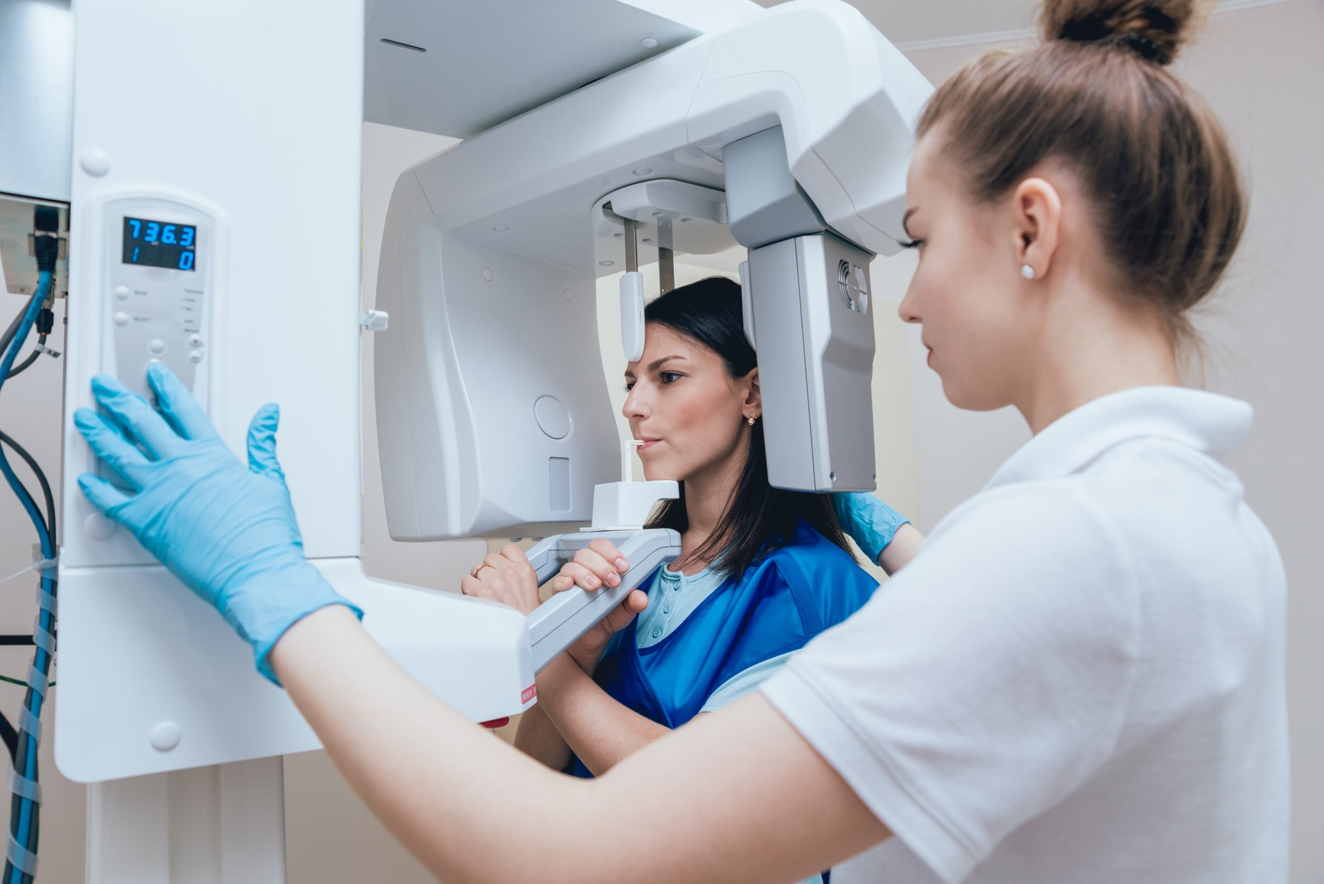
CBCT Machine
Dental cone beam computed tomography (CBCT) is a special type of x-ray machine that is implemented in scenarios where normal dental or facial x-rays are insufficient. This variation of the CT scanner employs a special type of technology to generate 3-D images of dental structures, soft tissues, nerve paths, and bone in the craniofacial area, in one scan. These images allow for more specific treatment planning. The CBCT machine has an x-ray beam, in the shape of a cone, which moves around you to create a large number of high-quality images, or views. It was developed as a means to produce similar images to what a CT provides, though with a significantly smaller and less costly machine that could be situated in an outpatient office. Providing detailed images of the bone, the CBCT machine evaluates diseases of the jaw, dentition, bony structures of the face, sinuses, and nasal cavity. One shortcoming is that it does not provide the comprehensive diagnostic information available with conventional CT, especially in the analyzing of soft tissue structures such as muscles, glands, nerves, and lymph nodes. The CBCT machine can also be used for reconstructive surgery, cephalometric analysis, locating the origin of pathology, surgical planning for impacted teeth, diagnosing temporomandibular joint disorder (TMD), and for the accurate placement of dental implants.
-
Dental Lasers
Diode Laser | The diode laser is useful for procedures that involve soft tissues, and are great for sterilizing endodontic canals, treating periodontal disease, and teeth whitening. It is a tool that offers a wide array of clinical treatment possibilities and is capable of great precision thanks to its portability and touchscreen controls. It has been shown to be helpful in treating challenging periodontal conditions while providing rapid healing and reduced swelling.
BIOLASE Epic X Laser | BIOLASE Epic X is a state-of-the-art and well-conceived example of a dental diode laser. A variety of pre-set procedures are installed into the device, with suggested settings that can be accessed swiftly and easily by our dentist. The handpiece is fitted with several single-use tips to access any area of our mouths, and there is a handpiece to be used for bio stimulation in pain relief and a whitening contour handpiece for patients who wish to receive laser-assisted in-office whitening. Most commonly, the tips of preference are the ones used intraorally since they are disposable and adjustable to any angle.
-
Air-Filtration (Surgically Clean Air)
We understand that staying inside can cause air to become dense. As more people filter through an area, it is common that the air quality may lack in cleanliness. Due to the constant change in temperature, a building may hold onto bacteria, mold, or illnesses. However, we have an air-filtration system, commonly known as Surgically Clean Air. It is an air cleaning process that has a six-step filtration which helps remove any harmful bacteria in the surrounding area. Our patient's health is of the utmost importance to us, which is why we have implemented this system in our office.
-
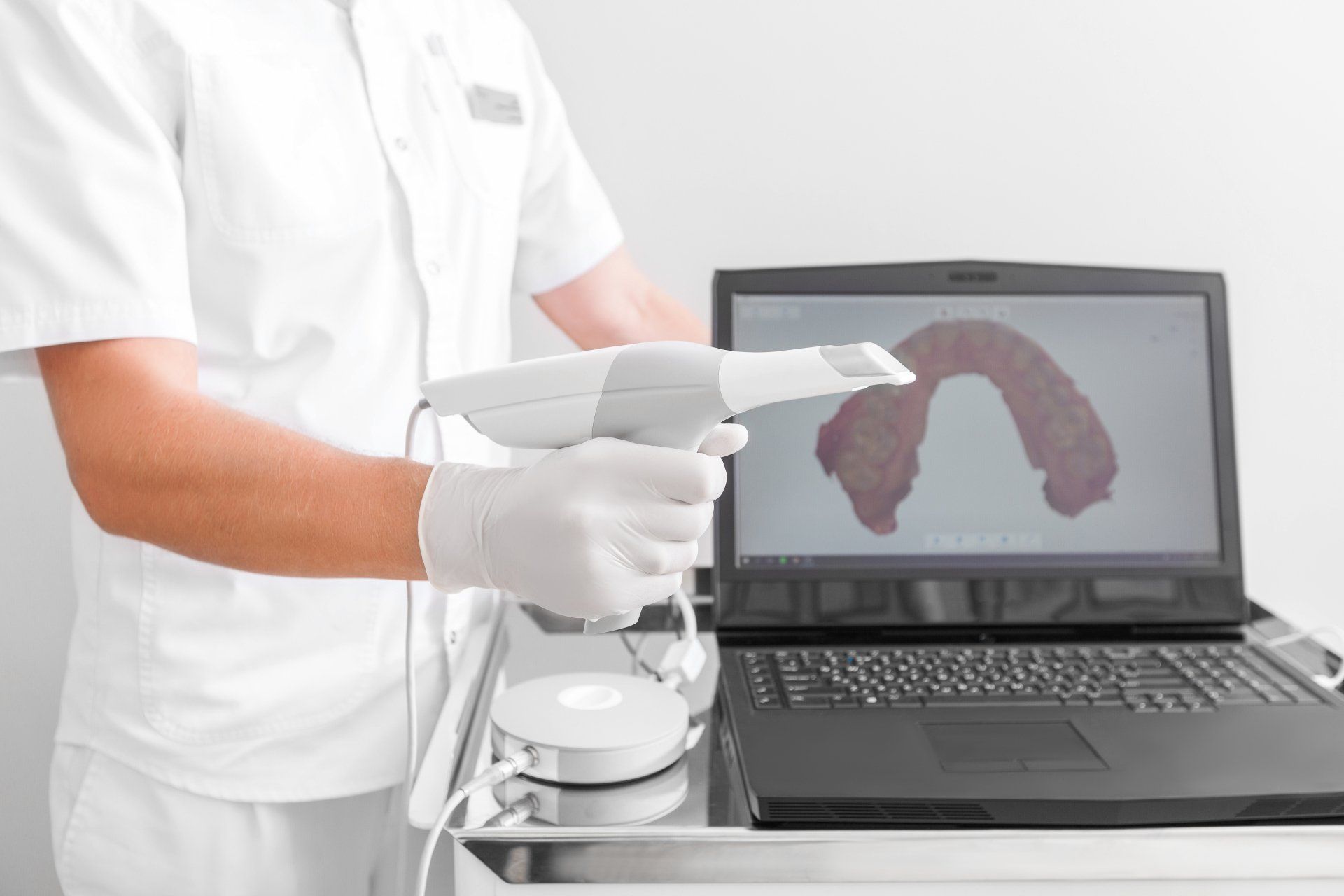
Intraoral Camera
About the same size as a marker, intraoral cameras are digital imaging tools used to create high-resolution images of your teeth, gums, and other hard-to-reach places in your mouth. Intraoral cameras help dental professionals detect dental issues, such as tooth decay, periodontal disease, and oral cancer. Other great benefits include:
- You can see, with precision, where you need to focus on brushing or flossing.
- You can see the difference before and after treatment.
- You can see magnified images of your teeth and gums, which helps dental professionals diagnose gum disease and cavities, and if caught early, can help prevent them.
- These photos provide proof for insurance companies to give you the coverage you need.
- Intraoral cameras also limit your time in the office because the images are produced in real-time, and the outcomes are available almost immediately.